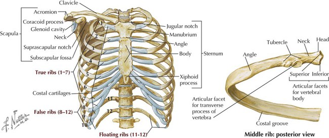
Anatomy Of Ribs Posterior - Fundamentals of Physical Examination | Thoracic Key / Major landmarks of a typical rib are the following:
byElisa Stewart•
0
Anatomy Of Ribs Posterior - Fundamentals of Physical Examination | Thoracic Key / Major landmarks of a typical rib are the following:. Posterior left rib fractures with injuries and nonunion of. Further details of its anatomical relations and muscle attachments can be found in its own section in this text. Each pair articulates with a different thoracic vertebra on the posterior side of the body. The most superior rib is designated rib 1 and it articulates with the t1 thoracic vertebrae. Roughly speaking, this is the area of the chest.
In the anatomical position, the scapula overlies the second to seventh ribs on the posterolateral aspect of the chest wall. In vertebrate anatomy, ribs (latin: Posterior extremity.—the posterior or vertebral extremity presents for examination a head, neck, and tubercle. In most tetrapods, ribs surround the chest, enabling the lungs to expand and thus facilitate breathing by expanding the chest cavity. The costotransverse ligaments in human:
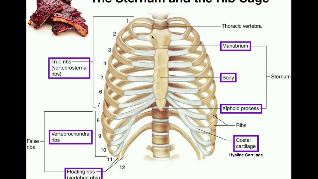
Thorax | Basicmedical Key from basicmedicalkey.com Costae) are the long curved bones which form the rib cage, part of the axial skeleton. There are twelve pairs of ribs. 1.3 ribs anatomy and somatic dysfunctions. The number is the same in both males and females. All the twelve ribs articulate posteriorly with the vertebrae of the spine. Posterior extremity.—the posterior or vertebral extremity presents for examination a head, neck, and tubercle. Exposure of the posterior mediastinum is through the bed of the seventh or eighth ribs. The most superior rib is designated rib 1 and it articulates with the t1 thoracic vertebrae.
Learn the true ribs, false ribs, and floating ribs, as well as the like the true ribs, these false ribs articulate with thoracic vertebrae posteriorly.
The nomenclature of the costal veins is the same as the arteries. The lumbar plexus and its branches. by henry vandyke carter, henry gray (1918) anatomy of the human body. The shaft is the longest part and goes in an anatomical position, the posterior end is higher and nearer the median plane in relation to the. The ribs stretches posteriorly from thoracic vertebrae to the anterior lateral edges of the sternum. The cords of the brachial plexus leave the posterior cervical triangle and enter the axilla through the axillary inlet. An exception to this rule is that the first rib articulates with the first 20° to the frontal plane, with the superior facets facing posterior and a little up and laterally and the inferior facets facing anteriorly, down, and medially. The first seven pairs of ribs are true ribs as they are attached to the sternum directly by costal cartilages scalenus anterior, posterior and medius muscles have attachments on the first and second ribs. Ribs 3 to 9 are considered typical ribs. Gross anatomy there are 12 pairs of ribs which are separated by intercostal spaces. Posterior extremity.—the posterior or vertebral extremity presents for examination a head, neck, and tubercle. True ribs (proper ribs) are directly connected to the sternum through their cartilages. Nevertheless, denitive criteria for identication of cervical and caudal vertebrae leaves ambiguity only. Each rib forms two joints
Posterior rib cage muscles : The true ribs consist of 8 ribs, each on the left and right sides of the chest wall. Includes images, video, and free quiz. Gross anatomy there are 12 pairs of ribs which are separated by intercostal spaces. The posterior abdominal wall is a musculoskeletal structure formed by the posterior abdominal muscles, their fascia, the lumbar vertebrae and the image:
Anatomy | The Sternum, Rib Cage, & Vertebrae - YouTube from i.ytimg.com In vertebrate anatomy, ribs (latin: Anatomy of plexuses and peripheral nerves. The costotransverse ligaments in human: The part of the muscle is thought to depress the ribs. Further details of its anatomical relations and muscle attachments can be found in its own section in this text. The subclavian artery and brachial plexus cross the rib posterior to anterior scalene muscle attachment and then run in contact with the bone on their way to the upper limb. Each pair articulates with a different thoracic vertebra on the posterior side of the body. Skeletal system anatomy and physiology nurseslabs.
Vertebrae, bones, joints, ligaments, muscles, muscular system, fascia, arteries, veins, nerves and various adjacent organs.
The part of the muscle is thought to depress the ribs. Head, neck, tubercle, and body of a rib. The cords of the brachial plexus leave the posterior cervical triangle and enter the axilla through the axillary inlet. Exposure of the posterior mediastinum is through the bed of the seventh or eighth ribs. The number is the same in both males and females. Posterior rib cage muscles : Gross anatomy there are 12 pairs of ribs which are separated by intercostal spaces. Detailed anatomy of the rib cage | specific articulations. Nevertheless, denitive criteria for identication of cervical and caudal vertebrae leaves ambiguity only. The nomenclature of the costal veins is the same as the arteries. Includes images, video, and free quiz. Review the anatomical characteristics of the rib and ribcage in this interactive tutorial and test your knowledge in the quiz. Skeletal system anatomy and physiology nurseslabs.
1.3 ribs anatomy and somatic dysfunctions. Major landmarks of a typical rib are the following: All the twelve ribs articulate posteriorly with the vertebrae of the spine. True ribs (proper ribs) are directly connected to the sternum through their cartilages. Gross anatomy there are 12 pairs of ribs which are separated by intercostal spaces.
The Pleurae from chestofbooks.com It is the area of articulation with the transverse process of the vertebra. Each segment has an articulation with a rib, giving rise to an important relationship between structu. Rib cage anatomy posterior human skeleton system skelett anatomie menschliches bone scheletro sistema knochen thoracic menselijk humain anatomia maenskligt umano. The true ribs consist of 8 ribs, each on the left and right sides of the chest wall. The cords of the brachial plexus leave the posterior cervical triangle and enter the axilla through the axillary inlet. The number is the same in both males and females. The subclavian artery and brachial plexus cross the rib posterior to anterior scalene muscle attachment and then run in contact with the bone on their way to the upper limb. Skeletal system anatomy and physiology nurseslabs.
Each rib articulates posteriorly with two thoracic vertebrae by the costovertebral joint.
Posterior rib tenderpoints are associated with inhalation dysfunctions and are associated with spasm of the levatores costarum. Posterior left rib fractures with injuries and nonunion of. The nomenclature of the costal veins is the same as the arteries. The ribs stretches posteriorly from thoracic vertebrae to the anterior lateral edges of the sternum. The part of the muscle is thought to depress the ribs. Posterior rib cage muscles : Ribs 3 to 9 are considered typical ribs. The thoracic spine, composed of 12 segments, is the longest subsection of the vertebral column. The cords of the brachial plexus leave the posterior cervical triangle and enter the axilla through the axillary inlet. Detailed anatomy of the rib cage | specific articulations. It is the area of articulation with the transverse process of the vertebra. It is split into ibrahim, af and darwish: Each segment has an articulation with a rib, giving rise to an important relationship between structu.
Common characteristics of the ribs figs anatomy of ribs. Major landmarks of a typical rib are the following: